Michigan Head & Spine Institute, Author at Michigan Head & Spine Institute Blog - Page 2 of 8

MHSI_web-1319103349-1200x683.jpg


Dr. Fredrick Junn
Fredrick S. Junn, M.D.
Neurosurgeon

While most people have heard of Parkinson’s disease, which affects nearly 1 million people in the United States, fewer of us are aware of its symptoms, risk factors and treatment.

Knowing about Parkinson’s Disease, including early signs to watch for, can help you and your loved ones catch the disease earlier, understand its signs, and learn how to manage it.

What Is Parkinson’s Disease?

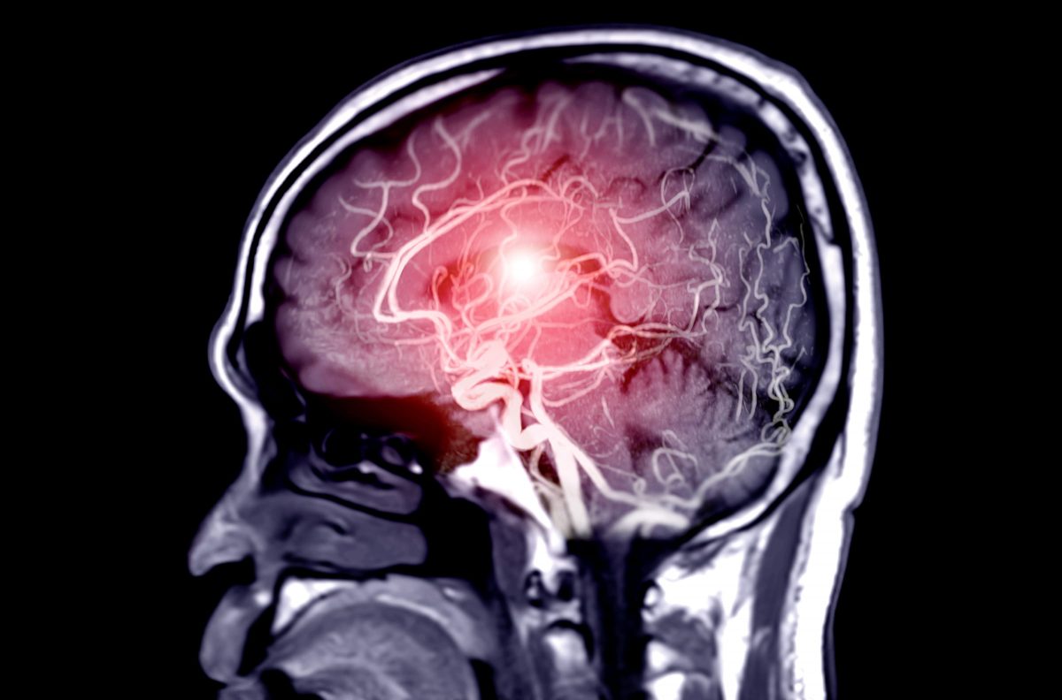
Parkinson’s disease is a disorder of the central nervous system that affects movement, specifically how your brain communications with the rest of your body to create and execute movement.

With Parkinson’s disease, damage to nerve cells in the brain lead to reduced levels of dopamine, which plays a role in movement. When dopamine decreases, the brain and the body can’t communicate properly, leading to symptoms of Parkinson’s disease.

Parkinson’s disease is a progressive neurological condition, meaning it starts slowly and gets worse over time. It typically starts in one portion of the body, such as the arm or leg, and eventually crosses over to affect the other side of the body as well. Early- to mid-stage symptoms of Parkinson’s disease include:

  • Tremors/shaking in fingers, thumb, hand or chin
  • Stiffness
  • Constipation
  • Trouble sleeping
  • Smaller handwriting (micrographia)
  • Speaking in a lower or softer voice (hypophonia)
  • Dizziness
  • Slowed movement (bradykinesia)
  • Loss of smell
  • Stooped posture

Very recently, new research has identified two more early signs of Parkinson’s disease: hearing loss and epilepsy.

Since many of these symptoms could be caused by other health conditions, it’s important to talk with your healthcare provider to potentially rule out Parkinson’s disease if you’re experiencing them.

As Parkinson’s disease progresses, so do the symptoms. Some that may show up later in the course of the disease include:

  • Tremors and stiffness on both sides of the body
  • Walking problems
  • Loss of balance
  • Shuffling gait
  • Markedly stooped posture

Parkinson’s disease can also affect a person’s mental health, and is linked to conditions such as depression, anxiety and dementia.

Risk Factors for Parkinson’s Disease

Although scientists haven’t been able to pinpoint what causes Parkinson’s disease, they have identified some risk factors. These include:

  • Genetics
    When people develop Parkinson’s disease at a young age (in their 20s or 30s), the cause is often genetic.
  • Advanced age
    The average age of onset for Parkinson’s disease is around 60 years old.
  • Gender
    Parkinson’s disease is more common in men than women.
  • Environmental triggers
    Exposure to pesticides/herbicides, heavy metals and other toxic chemicals has been shown to increase the risk for developing Parkinson’s disease. A street drug called “synthetic heroin” is also associated with Parkinson’s disease.

Treatments and Management for Parkinson’s Disease

There is no cure for Parkinson’s disease, but it can be managed and even slowed, depending how early the disease is diagnosed. Treating Parkinson’s disease typically involves increasing the amount of dopamine in the brain. This can be done with medications, including some types of antidepressants; newer anti-Parkinsonian medications are designed to provide a steady level of dopamine so patients don’t experience the crash associated with earlier treatments.

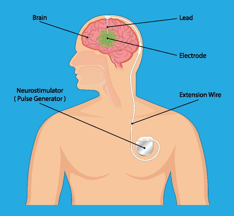
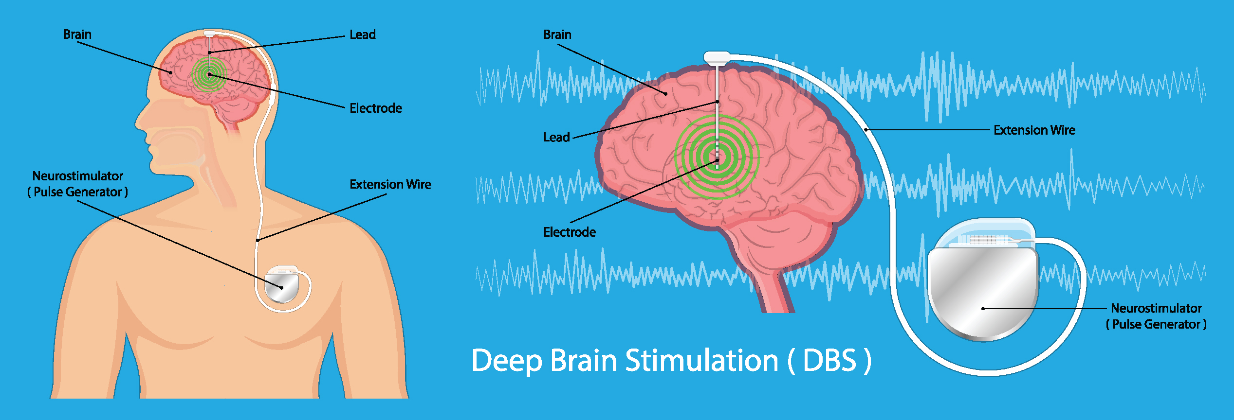
In addition to medication, deep brain stimulation (DBS) can be helpful for managing Parkinson’s disease symptoms. DBS is a surgical procedure where electrodes are implanted that deliver tiny electrical impulses to the parts of the brain that control movement. A major advantage of DBS is that it levels off dopamine levels without the side effects associated with some Parkinson’s disease medications.

Keeping the body healthy and moving are also key to controlling Parkinson’s disease symptoms, and many people find physical therapy to be helpful for maintaining movement.

If you think you may be experiencing Parkinson’s disease symptoms, or if you have a family history of the disease, you may want to ask your healthcare provider for a neurological referral. A neurologist can provide a diagnosis and start you down the path of managing your Parkinson’s disease.


If you or a loved one is seeking treatment for a condition of the brain, or to schedule an appointment with Dr. Junn or any of our MHSI experts, visit www.mhsi.com/appointments or call 248-784-3667.

MHSI-1248544072_aneurysm-1200x792.jpg


Dr. John Whapham
John Whapham, M.D., MS, FSNIS, FAAN
Neurosurgeon

Surgery has always been an option for certain types of intracranial cerebral aneurysms, but in recent years, minimally invasive procedures have greatly reduced both physical trauma as well as recovery times.

Cerebral aneurysms occur when part of a blood vessel wall weakens, allowing it to bulge, widen and potentially burst or rupture, which can be life-threatening.

Aneurysms can exist for years without detection. Because of this, there are often no symptoms until an aneurysm ruptures. Once that happens, symptoms may develop suddenly and violently, depending on the location of the aneurysm.

Usually, symptoms come on quickly and are unlike anything you might have experienced before. If you don’t often get headaches but suddenly experience an intense migraine, a ruptured brain aneurysm could be the culprit.

Call 911 and seek immediate medical attention if you or someone you know is having aneurysm signs and symptoms including:

  • Severe headache
  • worst headache of life
  • Stiffness or swelling in the neck
  • loss of consciousness
  • speech difficulty
  • focal motor or sensory symptoms weakness or numbness
  • Dizziness
  • Vision changes
  • Confusion
  • Hoarseness
  • Difficulty swallowing
  • Nausea and vomiting
  • Shock (low blood pressure, rapid heart rate, clammy skin, decreased awareness)

Sometimes an unruptured aneurysm is found when performing unrelated diagnostic testing such as MRIs or CAT scans.

Minimally Invasive Aneurysm Surgery Saves Lives

Aneurysm treatment has greatly improved in the last 25 to 30 years. In the past, major surgery, such as open craniotomy, was commonplace. In this procedure, a small metal clip is placed across the base of the aneurysm bulge that seals off blood flow to the ballooned blood vessel.

However, modern advances have made this major procedure less common.

These days, minimally invasive procedures such as coiling and stenting are the norm. Both divert the flow of blood away from the aneurysm making it less likely to rupture and are performed without opening the skull.

  • Coiling, also called endovascular embolization, uses a catheter passed through the groin up into the artery containing the aneurysm where tiny platinum coils are released. The coils induce clotting of the aneurysm and that clotting prevents blood from entering the aneurysm.
  • Stenting is when a catheter is used to place a stent (a soft, flexible mesh tube) into the blood vessel where an aneurysm has formed. The stent prevents blood from entering the aneurysm. In time, new cells grow on the stent, sealing the aneurysm and healing the vessel.

These minimally invasive procedures allow for a much shorter recovery period, and it is possible to return to daily living as early as one day after hospital discharge.

In fact, some patients with an unruptured aneurysm check in at the hospital, undergo a procedure that is finished in a couple of hours, stay overnight for observation and are discharged the next morning with only a small bandage covering the spot in the groin or wrist where the needle access was made.

Quick Treatment Is Key

It’s crucial to contact first responders at the first sign of an intense, sudden headache since that may indicate a ruptured aneurysm.

If you are diagnosed with an unruptured aneurysm, call us right away to make an appointment.

Depending on the aneurysm’s size and location, you may require regular checkups from a physician trained in aneurysm treatment and surgery. A treatment plan, including lowering your blood pressure, quitting smoking and medical intervention, can help you reduce the risk of rupture.


If you or a loved one is seeking treatment for a condition of the brain, or to schedule an appointment with Dr. Whapham or any of our MHSI experts, visit www.mhsi.com/appointments or call 248-784-3667.

MHSI-Telehealth-1286810754-1200x800.jpg

Dr. Richard D. Fessler
Richard D. Fessler , M.D. Neurosurgeon

In 2013 Dr. Richard Fessler predicted the use of telehealth would become the norm as technology was in place to create visits for patients via a smart phone. Fast forward to 2020, Dr. Fessler saw his prediction become reality. The pandemic of a lifetime gave the entire healthcare industry a push into telehealth. Insurance companies expanded coverage for this type of visit with a push from Center for Medicare Service or CMS to cover costs.

“At MHSI we were ready to offer our patients this option when COVID caused concerns about safety for our patients and staff.  With the insurance barrier removed, it brought reassurance we could deliver the telehealth option and patients didn’t have to be concerned about unnecessary out-of-pocket-costs,” said Dr. Fessler. “We are thankful that our patients accepted the technology and the new way we are able to provide care.”

For example, when Tony couldn’t take his back pain any longer, he met with Neurosurgeon Dr. Ratnesh Mehra virtually and had trust that his surgical experience would be equally the same and relieve his back pain. Dr. Mehra recalls the day they met online, “Tony was able to effectively communicate with me about his pain and what he was feeling. At that point we decided that surgery for Tony was the right course of treatment and I met Tony in pre-op in person for the first time.” Tony says, “One week after surgery I was walking…dancing.”


Watch how intense pain stopped Tony in his tracks!:


To schedule a telehealth appointment with an MHSI neurosurgeon, call 248-784-3667 or visit MHSI.us.

Nasal-Endoscopy_Steven_thumb-1200x772.jpg

Every year neurosurgeons advance the procedures performed to treat brain and spine conditions. Just a few short years ago minimally invasive surgery of any type was unheard of. Yet today, an incision of less than 2 inches allows a neurosurgeon to fix or replace a disc in the neck or spine.

Minimally invasive nasal endoscopy is yet another advancement to treat tumors of the brain. This approach allows the neurosurgeon to access the brain to remove a tumor without performing a craniotomy, which makes a “window” in the bone to allow access to the brain.

Dr. Jeffrey T. Jacob
Jeffrey T. Jacob, M.D. Neurosurgeon

Deciding between a craniotomy or using the nasal cavity to reach the brain, depends on what is best for the patient. “The goal is to be able to reach the tumor and remove all of it,” says Jeffrey Jacob, M.D. “Often, we are able to do that going through the nose with an endoscope, with a very small camera device.”

There are many types of brain tumors that can be treated with this approach. Brain tumors that are removed using the nasal endoscopic approach can be benign or malignant, and typically sit at the base of the skull or under the brain. This procedure removes the tumor and the blood supply to the tumor.

To perform a nasal endoscopy, Dr. Jacob partners with Adam Folbe, M.D., a rhinologist and endoscopic skull base surgeon at Michigan Sinus and Skull Base Center. Dr. Folbe goes through the nose and opens the window to the brain. He says, “There is no cutting of skin, muscle or fat resulting in no scars. Using this approach maximizes retrieval of the tumor with lesser disruption to the brain compared to the craniotomy approach.”

Patients who experience this approach have no signs of surgery on the outside of their body, because the nasal cavity is repaired with the patient’s own tissue, without stitches or sutures. During the post-surgical healing, patients are restricted from sneezing, blowing their nose, lifting and bending over for about three weeks.

Like any surgery, there can be side effects, like in Valerie’s case. Because of the location of her tumor, she lost her ability to smell. For Stephen, he experienced added restrictions as he waited for his vision to return to normal once the tumor was removed. Kevin didn’t experience difficulties after his surgery, but he did work hard to regain his strength and balance.

If you or a loved one is diagnosed with a brain tumor, consider all of your options and seek a second opinion. Like Valerie, you might find an option you didn’t expect.


Watch Stephen’s brain tumor story below:

Watch how a large pituitary tumor interrupted Kevin’s life:

Watch Valerie’s full story below:


To schedule an appointment with Dr. Jacob or any of the neurosurgeons at MHSI, call 248-784-3667 or visit MHSI.us

race47361-social1200x630.bzcJmm.jpg

September marks another effort to raise awareness about Chiari malformation. Chiari malformation is a disease of the brain where brain tissue is pushed down into the spinal canal. It occurs when the base of the skull is abnormally small or misshapen, causing pressure on the brain which forces it downward. Chiari malformation may cause blockage in the flow of the cerebrospinal fluid, which may cause the normal fluid spaces in the brain to enlarge, also known as hydrocephalus.

It was estimated that the condition occurs in about one in every 1,000 births. However, the increased use of diagnostic imaging has shown that Chiari malformation may be much more common. Complicating this estimation is the fact that some children who are born with the condition may not show symptoms until adolescence or adulthood, if ever. It can also be hereditary, and often those with Chiari will recall a family member with the same symptoms. Family members such as siblings Ezra and Katelyn, and mother Tammy and daughter Sheena.

Watch Ezra and Katelyn’s journey with Chiari below:

Chiari Malformation Corrected for Mother Tina and Daughter Sheena:

Adults and children alike can go through life and from doctor to doctor, with severe symptoms and are not diagnosed until an aware physician recognizes the symptoms. Patients Tina, Lu Anna and Tyler each had a physician acknowledge their symptoms and refer them to the leading expert in Chiari decompression, Neurosurgeon Dr. Holly Gilmer.

To learn more about Chiari malformation, the symptoms, and treatments, click here.

For most patients, the diagnosis is elusive for many years until the patient meets a doctor who is aware of Chiari, and understands the symptoms presented can become more manageable with surgery. One of the goals of Conquer Chiari Walk Across America is to raise awareness and funds to support further research. At this event, patients, their families and friends, along with doctors join in to walk to raise funds every third and fourth Saturday in September.

Click the graphic below to download the flyer:

Conquer Chiari Walk Across America


If you or a loved one is experiencing symptoms of Chiari malformation, call 248-784-3667 or visit MHSI.us to schedule an appointment with Dr. Gilmer.


BrainTumor-1250205787-1200x772.jpg

Medical illustration of brain tumor

It doesn’t get much more serious than being given the diagnosis that you have a brain tumor. Though brain cancer is rare and may be a life-threatening situation when detected, many forms of tumors can be successfully treated.

A brain tumor is a collection, or mass, of abnormal cells growing in, or next to the brain. But, what are the different types of brain tumors, and what are the symptoms to look out for?

Brain Tumor Symptoms
There are so many different types of brain tumors, and because each is so complex, brain tumors can cause different effects for each person. Physical problems and behavioral changes can occur throughout the body severely impacting one's life. The symptoms will depend on the individual's particular tumor, its location, and can result in:

  • Drowsiness
  • Personality changes
  • Confusion
  • Impulsiveness
  • Blurred vision
  • Balance problems
  • Headaches
  • Nausea
  • Vomiting
  • Seizures

Types of Treatable Brain Tumors
Though there are many different types, a brain tumor is classified by where it was originally formed. If it originated in the brain, it’s called a primary tumor and can be either noncancerous (benign), or cancerous (malignant). A tumor that originates in another part of the body and travels to the brain is called a meta tumor, and they are always cancerous.

Metastatic Brain Tumors - Also known as secondary tumors, they originate outside of the brain in another part of the body and then spread to the brain.

Meningiomas - Originating in the meninges, these thin layers of protective tissues surround the brain and spinal cord. Meningiomas usually grow slowly and most are not cancerous.

Pituitary Tumors - Found just under the brain, the pea-sized pituitary gland makes hormones that affect many of the body’s functions. A pituitary tumor can cause it to release too much, or too little, of these hormones which can cause serious problems.

Glioblastoma Multiforme - These are the most challenging forms of brain tumors to treat. MHSI physicians have access to the most current technology to deal with this cancer including image-guided surgical treatments.

Schwannoma - The most common type of benign peripheral nerve tumor in adults and rarely cancerous. When schwannoma grows larger, it can make removal more difficult. This type of nerve tumor can occur in any part of the body, and at any age.

Keith’s Brain Tumor Story
Before Keith, a patient at Michigan Head and Spine Institute, was diagnosed with his brain tumor he was an accomplished musician hoping for a big break.

“I got my first guitar when I was 12. It’s what calms me. It’s where I can focus on one thing and the thousand thoughts going on in my head will go away.”

But, that was all about to change when he started experiencing unexplained health problems and physical ailments.

“The first symptoms I had were the nausea, dizziness and headaches,” he said, “It got to the point where I couldn’t eat. The only thing that was staying down was water.”

Knowing something was wrong with his health, he went to an emergency room hoping it was just an ear infection. After undergoing some tests, his ER doctor came back with unsettling information.

“A brain tumor was not on my list.” - Keith, a patient at MHSI

Diagnosed with a sporadic case of hemangioblastoma, a benign tumor, Keith was seen by Michigan Head and Spine Institute, trusted experts for treatment and diagnosis of tumors, diseases and other conditions of the brain.

After reviewing his CT scan, it was confirmed Keith had a large mass at the back part of the head in the region of the cerebellum. This part of the brain also contributes to coordination.

“I just wanted it to stop hurting, to stop feeling sick,” Keith recalled.

While being prepped for surgery he also remembered the last thing he told the anesthesiologist, “I said, ‘I’m a musician.’ When I came back out, I still wanted to be a musician.”

Keith’s surgery was successful. He then underwent a second nonsurgical procedure called Gamma knife treatment to remove the last bit of his tumor.

“When I woke up after the surgery, I knew it was fixed. The pain I was having before was gone,” he said relieved, “I like the way my doctor had a very confident way about himself.”

As for Keith’s guitar playing and music career after his surgery at MHSI:

“I’ve been in a few bands, had a little stardom,” he updated, “I’ve met a lot of people in the industry since the surgery that say they’re going to help me, so we’ll see what happens.”

To learn more about conditions of the brain and brain tumor procedures, please visit our MHSI Patient Education page at: Conditions of the Brain.

If you or a loved one is seeking treatment for a condition of the brain, or are seeking a second opinion on an existing diagnosis, schedule an appointment online or contact MHSI at 248-784-3667.

To view more information about MHSI’s neurosurgeons, visit: https://www.mhsi.com/doctors/neurosurgery-physicians/. For more information about MHSI’s  neurologists, visit: https://www.mhsi.com/doctors/neurologists/


2ndOpinion-1270585289-1200x800.jpg

The Importance of Getting a Second Opinion and Gaining Peace of Mind
If your body is telling you one thing, and your physician another, it may be time to seek a second opinion regarding surgery or treatment. When uncertain of an original diagnosis, or continuing to experience pain or discomfort – a second opinion can allow you to take control of your health and put your mind at rest.

Why Should You Get a Second Opinion?
The Mayo Clinic conducted a study of 286 patients that found as many as 88% of the people seeking a second opinion will leave a physicianʼs office with a new or redefined diagnosis. Of those patients, 21% will leave with a “distinctly different” diagnosis. Whereas only 12% will learn that the original diagnosis was correct. These results showed that one out of every five patients was incorrectly diagnosed.* 

Percentages such as these prove the importance of looking for a second opinion when necessary, and that patients should follow to their instincts.

When You Should Get a Second Opinion?
If you are not getting the answers that help you make an informed decision regarding a surgical procedure, or treatment option, you should ask more questions of your doctor. Ask yourself if pursuing the diagnosis will have lifelong consequences to your lifestyle and wellbeing. Would it be invasive or a risk to your quality of life? Focus on chronic and serious conditions when questioning treatment outcomes, and when inquiring about exploring other treatment options.

A physician should accept and respect your wishes for seeking a second opinion. They can also assist by offering referrals, and providing all of your medical records and test results. This will help your physician to communicate with the second physician, if needed.

Watch Valerie’s full story below:

Angela’s Second Opinion Story
“I was hit by a drunk driver, and immediately had this pain down my arm and wrist,” Angela said.

She’d gone to her primary care physician who diagnosed her injury as a pulled muscle from the seat belt. After seeing a chiropractor, and a neurologist, her pain still continued to worsen. Frustrated, Angela knew she required a second opinion.

“I was devastated and couldn’t do this anymore,” she explained, “My husband said to phone our insurance company and have them recommend another doctor for me, or we were going to find one.”

Angela’s insurance adjuster gave her the phone number for Michigan Head and Spine Institute.

“From the very first appointment I had a new diagnosis, a plan of action and was set up with a physician for pain management,” Angela said, finally relieved, “This doctor literally saved my life. Between his pain management and conservative treatment, Michigan Head and Spine Institute went above and beyond.”

Watch Angela’s full story below:

How a Patient Can Get a Second Opinion 
Patients have rights – you have the right to a second opinion. MHSIʼs multidisciplinary team can offer further knowledge and peace of mind for a brain, neck or spine diagnosis or surgery. If you, or a loved one, need more information about getting a second opinion contact Michigan Head and Spine Institute at 248-784-3667 or visit MHSI.us to schedule an appointment.

Immediate Telehealth Appointments Available.

*Mayo Clinic, 2017. Journal of Evaluation in Clinical Practice


DBS_Thumbnanail.jpg

Movement disorders refer to conditions which cause abnormal voluntary or involuntary movements. Symptoms are caused by disorganized electrical signals in specific areas of the brain. Those challenged with essential tremor, Parkinson’s disease, dystonia, and other neurological conditions may benefit from deep brain stimulation (DBS), especially when symptoms progress or medications become less effective.

DBS is performed by neurosurgeons who precisely place one or more wires, called electrodes or leads, inside the brain. The lead is connected to a pacemaker-like device called a neurostimulator that is surgically implanted into the patient’s chest. The neurostimulator then conducts continuous pulses of electric current through the leads to interrupt the disorganized brain signals causing tremor or other movement disorder symptoms.

Insertion of the leads and neurostimulator are typically accomplished in two steps. First, the neurosurgeon places the leads either into one or both sides of the brain, and this is often performed while the patient is awake. In a second procedure the neurostimulator is implanted in the chest while the patient is asleep. Programming of the neurostimulator can be performed by the neurosurgeon, neurologist, or primary care physician, to find the optimum settings that are effective for each patient. There are different devices available that can tailor therapy to each individual patient, and the device representative often has an important role to help the patient become familiar with their programmer and programming settings.

For patients with essential tremor, the most common movement disorder, DBS can return a person to normal daily activities like dressing, shaving, eating, and drinking. Symptoms of Parkinson’s disease such as tremor, bradykinesia and rigidity are especially well-treated with DBS, and medication dosages can often be decreased. Dystonia is an uncommon movement disorder, with symptoms of abnormal posturing and twisting movements, which also respond to DBS.

DBS can also be used to treat patients with epilepsy, obsessive-compulsive disorder, or certain pain disorders. These unique disorders are often managed by specialized neurologists and psychiatrists, and a thorough discussion with your specialist is necessary to determine if you are a candidate for this therapy.

At MHSI, a neurosurgeon or functional neurosurgeon will work with a patient’s neurologist or primary care physician prior to and after surgery to ensure that the optimal therapy is achieved.

For more information about DBS, visit our patient education page on the topic, click here. If you or a family member or friend might benefit from DBS, please schedule a consultation by calling 248-784-3667 or visit MHSI.us.

MHSI Neurosurgeons Who Perform DBS 

Dr. Fredrick Junn
Dr. Richard Veyna


TeleMed4-1227122484-1200x800.png

Michigan Head & Spine offers Telehealth appointments through Zoom Cloud Meetings. The following tips will help you prepare for a Telehealth visit.

Schedule an appointment
Your appointment can be scheduled through the office where you see your MHSI provider. On the day of your appointment you will receive a Zoom link and passcode. If you need to reschedule your appointment, please contact us within 24 hours of the appointment via the patient portal or by calling 248-784-3667.

Find the right location
Find a quiet place to communicate with your physician. Make sure this is a location where you won’t be disturbed by others coming into the room or background noises like the television, radio, pets, or noisy appliances. Be sure to have plenty of space around you – you may be asked to move around during your appointment so your physician can see your movements. DO NOT hold your Telehealth visit while you are driving or sitting in a vehicle – MHSI will reschedule your appointment as a result. Make sure there is plenty of light in the space where you hold the meeting so your provider can see you clearly.

Prepare your personal details
Come prepared to discuss your medical history or any pre-existing conditions you have. Be sure to mention any symptoms you may be experiencing since your last appointment. Check your prescriptions so you can discuss refills if they are needed. And, be sure to write down any questions or concerns you may have so you don’t forget to ask during the appointment. 

Set up technology
Telehealth appointments require an internet-connected device and use of a video source from a smartphone, computer with camera, or tablet. Make sure that your equipment is fully charged or plugged in and reliably connected to the internet before your appointment. Headphones or earbuds provide the best sound quality and can help reduce feedback or background noise.

Our virtual appointments are available through the Zoom Cloud Meeting application. Be sure that you have downloaded the most current program from the App store for your phone, tablet or computer. The application is free to download.

Test your software in advance of your scheduled appointment by visiting the Zoom website at https//zoom.us/test. 

Get connected
Be sure to be connected to the internet and locate the meeting link. Follow these steps to connect:

  • Click the Zoom link in the email or text to launch the Zoom application. 
  • Enter the Meeting ID and Passcode from the email appointment to join the meeting. 
  • You will see a preview window and a button that states “Join with Video,” click the button to begin. 
  • You may be asked by Zoom to allow permissions or enable video and audio.
  • Join the audio by selecting “Call via device audio” or “Call via internet”
  • Please be patient with your provider if they do not arrive precisely at the appointment time.

For more assistance a short video on How to Join a Zoom Meeting can be viewed here.

We will try to assist you in getting connected the best we can – but we cannot provide IT support. Please have assistance available with you at your appointment time if you have difficulties connecting. If you are unable to get connected, your appointment may be rescheduled.

Click here to download our connection tips sheet and keep it for future reference during your scheduled appointment.