Brain Archives - Page 2 of 3 - Michigan Head & Spine Institute Blog

BrainTumor-1250205787-1200x772.jpg

Medical illustration of brain tumor

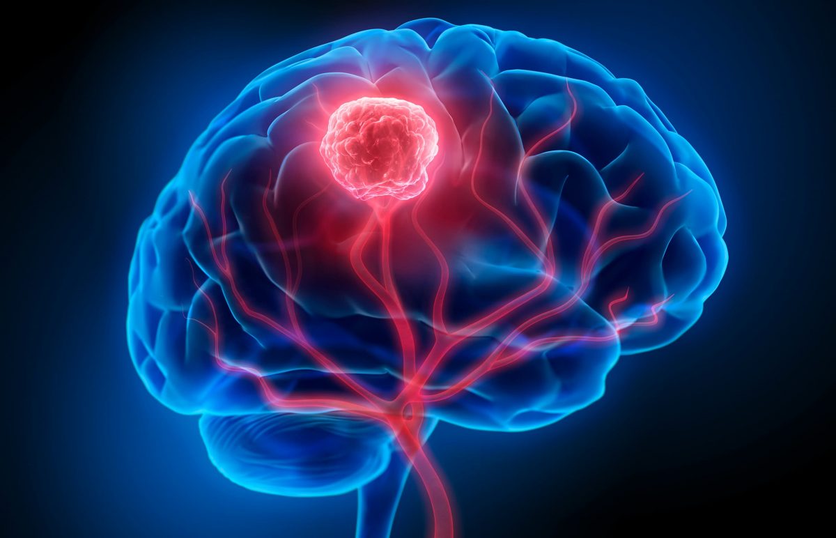
It doesn’t get much more serious than being given the diagnosis that you have a brain tumor. Though brain cancer is rare and may be a life-threatening situation when detected, many forms of tumors can be successfully treated.

A brain tumor is a collection, or mass, of abnormal cells growing in, or next to the brain. But, what are the different types of brain tumors, and what are the symptoms to look out for?

Brain Tumor Symptoms
There are so many different types of brain tumors, and because each is so complex, brain tumors can cause different effects for each person. Physical problems and behavioral changes can occur throughout the body severely impacting one's life. The symptoms will depend on the individual's particular tumor, its location, and can result in:

  • Drowsiness
  • Personality changes
  • Confusion
  • Impulsiveness
  • Blurred vision
  • Balance problems
  • Headaches
  • Nausea
  • Vomiting
  • Seizures

Types of Treatable Brain Tumors
Though there are many different types, a brain tumor is classified by where it was originally formed. If it originated in the brain, it’s called a primary tumor and can be either noncancerous (benign), or cancerous (malignant). A tumor that originates in another part of the body and travels to the brain is called a meta tumor, and they are always cancerous.

Metastatic Brain Tumors - Also known as secondary tumors, they originate outside of the brain in another part of the body and then spread to the brain.

Meningiomas - Originating in the meninges, these thin layers of protective tissues surround the brain and spinal cord. Meningiomas usually grow slowly and most are not cancerous.

Pituitary Tumors - Found just under the brain, the pea-sized pituitary gland makes hormones that affect many of the body’s functions. A pituitary tumor can cause it to release too much, or too little, of these hormones which can cause serious problems.

Glioblastoma Multiforme - These are the most challenging forms of brain tumors to treat. MHSI physicians have access to the most current technology to deal with this cancer including image-guided surgical treatments.

Schwannoma - The most common type of benign peripheral nerve tumor in adults and rarely cancerous. When schwannoma grows larger, it can make removal more difficult. This type of nerve tumor can occur in any part of the body, and at any age.

Keith’s Brain Tumor Story
Before Keith, a patient at Michigan Head and Spine Institute, was diagnosed with his brain tumor he was an accomplished musician hoping for a big break.

“I got my first guitar when I was 12. It’s what calms me. It’s where I can focus on one thing and the thousand thoughts going on in my head will go away.”

But, that was all about to change when he started experiencing unexplained health problems and physical ailments.

“The first symptoms I had were the nausea, dizziness and headaches,” he said, “It got to the point where I couldn’t eat. The only thing that was staying down was water.”

Knowing something was wrong with his health, he went to an emergency room hoping it was just an ear infection. After undergoing some tests, his ER doctor came back with unsettling information.

“A brain tumor was not on my list.” - Keith, a patient at MHSI

Diagnosed with a sporadic case of hemangioblastoma, a benign tumor, Keith was seen by Michigan Head and Spine Institute, trusted experts for treatment and diagnosis of tumors, diseases and other conditions of the brain.

After reviewing his CT scan, it was confirmed Keith had a large mass at the back part of the head in the region of the cerebellum. This part of the brain also contributes to coordination.

“I just wanted it to stop hurting, to stop feeling sick,” Keith recalled.

While being prepped for surgery he also remembered the last thing he told the anesthesiologist, “I said, ‘I’m a musician.’ When I came back out, I still wanted to be a musician.”

Keith’s surgery was successful. He then underwent a second nonsurgical procedure called Gamma knife treatment to remove the last bit of his tumor.

“When I woke up after the surgery, I knew it was fixed. The pain I was having before was gone,” he said relieved, “I like the way my doctor had a very confident way about himself.”

As for Keith’s guitar playing and music career after his surgery at MHSI:

“I’ve been in a few bands, had a little stardom,” he updated, “I’ve met a lot of people in the industry since the surgery that say they’re going to help me, so we’ll see what happens.”

To learn more about conditions of the brain and brain tumor procedures, please visit our MHSI Patient Education page at: Conditions of the Brain.

If you or a loved one is seeking treatment for a condition of the brain, or are seeking a second opinion on an existing diagnosis, schedule an appointment online or contact MHSI at 248-784-3667.

To view more information about MHSI’s neurosurgeons, visit: https://www.mhsi.com/doctors/neurosurgery-physicians/. For more information about MHSI’s  neurologists, visit: https://www.mhsi.com/doctors/neurologists/


DBS_Thumbnanail.jpg

Movement disorders refer to conditions which cause abnormal voluntary or involuntary movements. Symptoms are caused by disorganized electrical signals in specific areas of the brain. Those challenged with essential tremor, Parkinson’s disease, dystonia, and other neurological conditions may benefit from deep brain stimulation (DBS), especially when symptoms progress or medications become less effective.

DBS is performed by neurosurgeons who precisely place one or more wires, called electrodes or leads, inside the brain. The lead is connected to a pacemaker-like device called a neurostimulator that is surgically implanted into the patient’s chest. The neurostimulator then conducts continuous pulses of electric current through the leads to interrupt the disorganized brain signals causing tremor or other movement disorder symptoms.

Insertion of the leads and neurostimulator are typically accomplished in two steps. First, the neurosurgeon places the leads either into one or both sides of the brain, and this is often performed while the patient is awake. In a second procedure the neurostimulator is implanted in the chest while the patient is asleep. Programming of the neurostimulator can be performed by the neurosurgeon, neurologist, or primary care physician, to find the optimum settings that are effective for each patient. There are different devices available that can tailor therapy to each individual patient, and the device representative often has an important role to help the patient become familiar with their programmer and programming settings.

For patients with essential tremor, the most common movement disorder, DBS can return a person to normal daily activities like dressing, shaving, eating, and drinking. Symptoms of Parkinson’s disease such as tremor, bradykinesia and rigidity are especially well-treated with DBS, and medication dosages can often be decreased. Dystonia is an uncommon movement disorder, with symptoms of abnormal posturing and twisting movements, which also respond to DBS.

DBS can also be used to treat patients with epilepsy, obsessive-compulsive disorder, or certain pain disorders. These unique disorders are often managed by specialized neurologists and psychiatrists, and a thorough discussion with your specialist is necessary to determine if you are a candidate for this therapy.

At MHSI, a neurosurgeon or functional neurosurgeon will work with a patient’s neurologist or primary care physician prior to and after surgery to ensure that the optimal therapy is achieved.

For more information about DBS, visit our patient education page on the topic, click here. If you or a family member or friend might benefit from DBS, please schedule a consultation by calling 248-784-3667 or visit MHSI.us.

MHSI Neurosurgeons Who Perform DBS 

Dr. Fredrick Junn
Dr. Richard Veyna


TrentonOfficeGraphic.jpg

Downriver Communities Now Have Neurosurgery Expertise

When looking for a neurosurgeon, you want someone you can trust with head, neck and spine conditions. Someone who has expertise with a good reputation. Someone close to home.

MHSI opened a new office, conveniently located on the campus of Beaumont Hospital, Trenton. Staffed by neurosurgeons Ratnesh Mehra, D.O., Pradeep Setty, D.O.  and Jeffrey Jacob, M.D., we offer the caliber of care and treatment you expect, only closer to home.

Neurosurgery is delicate, and you want the best, most experienced surgeons. The team at MHSI – Trenton has a complex set of skills including minimally invasive and robotic surgery, superior knowledge of brain tumors, and leading-edge experience with skull base and spine surgery.

Patients have long traveled to MHSI to be seen by specialists who provide the full spectrum of care for head, neck and spine conditions, because we provide a range of treatment and diagnosis options that are unparalleled in Southeast Michigan. And with the new Trenton office so close to home, receiving our care just got easier.

This is our tenth location and our first Downriver. Opening another office cements our mission to you: Provide the best, most experienced care. Spine, neck and head conditions are complex, and you want the best surgeon. With the team at MHSI working for you, helping you make the best decisions for your health, you know you have the professionals you want on your side.

The new location on Fort Street, on the campus of Beaumont Hospital, Trenton, opens up conveniences for patients as well. Imaging, testing, therapies and more are within arm’s length at the hospital, while the doctors you trust are right there with you.

We are accepting new patients, seeing post-op patients and continuing to provide care for those in the follow-up stage of care. You can call 248-784-3667 for an appointment, or request one online.

Expertise makes a difference, and the difference is MHSI.


Steve-with-DSS-looking-at-Optune-500x500.jpg

Neurosurgeons at Michigan Head and Spine Institute have begun using Optune for the treatment of newly diagnosed and recurrent glioblastoma (GBM). Richard Veyna, M.D., is certified to prescribe this wearable and portable medical device, the first FDA-approved therapy in more than a decade for newly diagnosed GBM.

“Our goal at Michigan Head & Spine Institute is to treat cancer patients with the latest, approved therapies available, so we welcome the opportunity to provide Optune as part of a combination treatment for those fighting GBM,” said Richard Veyna, M.D. “We want our patients to have the best possible quality of life. With Optune as a therapy patients are able to go about their daily activities with minimal disruption to their lives.”

For newly diagnosed patients, Optune is used with the chemotherapy temozolomide (TMZ) after surgery and radiation with TMZ. In a clinical trial, adding Optune to TMZ was proven to delay GBM tumor growth and extend survival in newly diagnosed patients compared with TMZ alone. For recurrent patients, it can be used alone when surgery and radiation treatment options have been exhausted. Optune is approved for the treatment of adult patients (22 years of age or older) with GBM. In a clinical trial, adding Optune to TMZ provided an unprecedented five-year survival advantage in patients with newly diagnosed GBM.

About Glioblastoma Multiforme

Glioblastoma, also called glioblastoma multiforme, or GBM, is the most aggressive type of primary brain tumor. While GBM is rare, it is the most common type of primary brain cancer in adults. Approximately 12,500 new cases of GBM or brain tumors that may progress to GBM are diagnosed in the United States each year. (Watch Pamela’s story)

How Optune WorksOptune-Device-500x500

Optune creates low-intensity electric fields—called Tumor Treating Fields (TTFields)—which potentially slow or stop cell division leading to cancer cell death. Because TTFields do not enter the bloodstream like a drug, they did not significantly increase TMZ-related side effects for newly diagnosed patients. In clinical trials the most common device related adverse events were scalp irritation from device use and headache.

For the treatment of recurrent GBM, Optune is indicated following histologically–or radiologically–confirmed recurrence in the supratentorial region of the brain after receiving chemotherapy. The device is intended to be used as a monotherapy, and is intended as an alternative to standard medical therapy for GBM after surgical and radiation options have been exhausted.

Guidelines for Use of Optune

Optune should not be used if the patient has an active implanted medical device, a skull defect (such as, missing bone with no replacement), or bullet fragments. Use of Optune with implanted electronic devices has not been tested and may theoretically lead to malfunctioning of the implanted device.

Use of Optune together with skull defects or bullet fragments has not been tested and may possibly lead to tissue damage or render Optune ineffective. Do not use Optune if you are known to be sensitive to conductive hydrogels. In this case, skin contact with the gel used with Optune may commonly cause increased redness and itching, and rarely may even lead to severe allergic reactions such as shock and respiratory failure.

Warnings and Precautions

Optune should only be used after receiving training from qualified personnel, such as your doctor, a nurse, or other medical personnel who have completed a training course given by Novocure™ (the device manufacturer).

Optune should not be used if the patient is pregnant, or thinks she might be pregnant or are trying to get pregnant. It is not known if Optune is safe or effective in these populations.

The most common (≥10%) adverse events involving Optune, in combination with temozolomide, were low blood platelet count, nausea, constipation, vomiting, fatigue, scalp irritation from device use, headache, convulsions, and depression.

The most common (≥10%) adverse events seen when using Optune alone were scalp irritation from device use and headache.

Scalp irritation from device use, headache, malaise, muscle twitching, fall and skin ulcer is considered an adverse reaction related to Optune when using the device alone.

Cautions:

All servicing procedures must be performed by qualified and trained personnel, like Dr. Veyna.

Do not use any parts that do not come with the Optune Treatment Kit, or that were not sent to you by the device manufacturer or given to you by your doctor.

Do not wet the device or transducer arrays.

If you have an underlying serious skin condition on the scalp, discuss with your doctor whether this may prevent or temporarily interfere with Optune treatment.


surgical-mask-docs-1024x682.jpg

Patients arrive at Michigan Head & Spine Institute with many different conditions and injuries.  We hear from our patients that learning about conditions other than what you might be experiencing often helps family members or friends. So this month the MHSI Health Education Series will discuss trigeminal neuralgia.

Robert Johnson, M.D., MHSI neurosurgeon explains, “Pressure or damage to the trigeminal nerve can cause malfunction of the nerve which leads to the pain in the face known as trigeminal neuralgia.” Trigeminal neuralgia usually affects more women than men, and those over 50 years of age. Doctors may use medications, and sometimes surgery to relieve the pain and release the pressure on the nerve.

Some Possible Causes:

  • Contact between a normal blood vessel and an artery/vein
  • Aging
  • Multiple Sclerosis or other movement disorders
  • Brain lesion
  • Stroke or facial trauma
  • Abnormal position of blood vessel related to the nerve

Symptoms of Trigeminal Neuralgia

There are many symptoms of trigeminal neuralgia which could include one of these patterns:

  • Extremely painful, sharp electric-like spasms that may last a few seconds to a few minutes.
  • Spontaneous attacks of pain while doing regular daily activities such as talking, brushing your teeth, or chewing.
  • Pain that is triggered by sounds, wind or touch.
  • Pain that affects one side of the face, rarely does it affect both sides of the face.
  • Constant aching or burning pain.
  • Pain is usually in the areas that are supplied by the trigeminal nerve: cheek, jaw, teeth, gums, or lips. Pain in the eyes and forehead are less common.
  • Attacks become more frequent and increase in intensity.
  • Pain, for an unknown reason, isn’t usually felt while sleeping. Knowing this may help physicians pinpoint if it could be a migraine or toothache rather than trigeminal neuralgia.
  • In atypical trigeminal neuralgia, a severe migraine in addition to the sharp electric like spasms may be present.

Treatment for Trigeminal Neuralgia

Medication, which may include muscle relaxers, anti-seizure drugs, and antidepressants to target the inflamed nerve, may be prescribed to those diagnosed with trigeminal neuralgia. In some cases, surgery may be needed to relieve the pressure that is causing the nerve disruption. These surgical options may include:

  • Microvascular decompression – relocating or removing a blood vessel that is in contact with the trigeminal nerve. This is done with a small incision behind the ear on the side of your pain
  • Tumor Removal
  • Gamma Knife Radiosurgery
  • Glycerol injection – the sterile glycerol damages the trigeminal nerve and blocks pain signals
  • Balloon compression

Robert Johnson, M.D., Jeffrey Jacob, M.D., and Daniel Michael, M.D., are all MHSI neurosurgeons who specialize in treating trigeminal neuralgia.  If you identified these symptoms as those you may be experiencing, please call MHSI for an appointment, 248-784-3667.


iStock-613682270-1024x683.jpg

The two main blood vessels in the neck that supply blood to the brain are called the carotid arteries. When these arteries narrow, blood flow to the brain is reduced. This is called carotid artery stenosis. The gradual buildup of fatty substances and cholesterol deposits is called plaque.  Plaque occurs as we age, engage in unhealthy lifestyles and don’t manage risk factors, like high cholesterol.

Often, there are often no symptoms until a stroke occurs. So it is important to seek regular physical exams. If your doctor hears an abnormal sound in these arteries a carotid duplex or Doppler ultrasound may be required to examine the blood flow and look for plaque or blood clots.

Medication may be prescribed if less than 50% of the artery is blocked. If more than a 70% blockage is present a carotid endarterectomy or a carotid angioplasty / stenting procedure can improve blood flow to the brain.  Both procedures are usually conducted by a neurosurgeon. In the carotid endarterectomy, an incision is made in the neck and the plaque and diseased portions of the artery are removed to increase blood flow to the brain.

Richard Fessler, M.D., an endovascular neurosurgeon at MHSI explains, “In a carotid angioplasty, a catheter is inserted into the groin, through the aorta (the main blood vessel of your heart) in an attempt to clear the blockage and open up the artery. Sometimes a stent is inserted into the artery to keep the artery open and the blood flowing.”

For all patients, “These are much less invasive procedures than open surgery for all patients, but especially elderly patients,” adds Dr. Fessler.

If you find yourself in an emergency situation, like having a stroke that requires a carotid endarterectomy or angioplasty, or your doctor says you need one of these procedures ask for an MHSI neurosurgeon. To schedule an appointment, call 248-784-3667 or online at MHSI.us


Chiari MalformationIn her early thirties, Amy experienced very bad and massive migraine headaches. At that time, she went to see a neurosurgeon and was diagnosed with Chiari I malformation. Amy was told she could have brain surgery, but it was an elective surgery. She was told if it’s not bothering you – you shouldn’t fix it. Fast forward to now at age 43.

Amy is a kindergarten teacher of 27 students in her classroom. She would be exhausted and fight to stay awake. Everyday would be a challenge because of fatigue, headaches, and continuous scratching herself to stay awake. Five minutes driving in the car would be very stressful for her and falling asleep at the wheel was becoming a serious issue for Amy.

Chiari malformations are structural defects that occur in the cerebellum, the part of the brain that controls coordination and muscle movement. Previous estimates were that malformations occur in about one in every 1,000 births, but increased use of diagnostic imaging indicates that the disorder may be more common than once thought.

What is Chiari Malformation?

Holly Gilmer, M.D., neurosurgeon and a leading expert in Chiari malformation explains that older children experience headaches, dizziness, ringing in the ears, and problems with vision. One of the most frequent presentations is scoliosis with none of these symptoms except infrequent headaches. Some children may not have noticeable symptoms until adolescence or adulthood. In teen and adult years, problems can include persistent headaches, neck pain, and weakness and/or numbness and tingling in the arms and legs.

Adult symptoms include neck pain, balance problems, muscle weakness, numbness or other abnormal feelings in the arms or legs, dizziness, vision problems, difficulty swallowing, ringing or buzzing in the ears, hearing loss, vomiting, insomnia, or headache made worse by coughing, laughing, or straining. Hand-eye coordination and fine motor skills may be affected. Symptoms can change over time depending on the build-up of cerebrospinal fluid and pressure on the brain, spinal cord, and nerves.

Dr. Gilmer says that “surgical treatment to correct the compression involves removing a portion of the skull and usually part of the C1 vertebra. The cerebellar tonsils are usually partially removed. We always open the covering of the brain (dura) and use an expansion graft to make the dura larger and give the brain more room to expand.”

“It only takes one person to change your life and I’m very grateful to Dr. Gilmer and Michigan Head & Spine Institute,” says Amy.

To refer a patient for diagnosis of Chiari malformation or evaluation for decompression surgery, call 248-784-3667.


oldercouple-1024x682.jpg

by Daniel B. Michael, M.D., Ph.D., Emeritus and Retired Neurosurgeon

Everyone forgets, from the day we are born until we die. Memory is one of the most important functions our brains provide. When we forget or cannot recall an event, person or thing it can be frustrating. When such memories can never be recalled it can be life threatening.  Alzheimer Disease (AD) is the progressive loss of certain types of memory due to specific degenerative changes in the brain. Twelve years following the diagnosis, over 95% of AD patients are dead. Alzheimer Disease is fatal and currently there is no effective treatment. In 2012 there were 5.4 million estimated cases of AD with approximately 36 million cases worldwide. The incidence is expected to quadruple by 2050. In 2012 US the cost to care for AD patients was estimated to be $200 Billion; in 2050 the cost will increase to an estimated, $1.2 Trillion, 70% paid for by Medicare. Unless effective treatments for AD can be found we face an unprecedented healthcare crisis. Dr. James Fontanesi, a noted radiation oncologist then on staff at William Beaumont Hospital (WBH), Royal Oak, MI, observed that relatively low doses of external radiation had been used to treat abnormal deposits of amyloid in parts of the body other than the brain. Amyloid deposition in the brain has been thought to play an important role in the development of AD. He hypothesized that radiation could be used to reduce amyloid in the brains of AD patients and lead to improved memory function. Over the past five years Dr. Fontanesi gathered a team of radiation oncologists, radiobiologists, and behavioral psychologists to design and carry out animal laboratory studies to see if radiation would reduce amyloid in the brain, improve memory, and if so by what mechanisms this treatment worked. Daniel Michael, M.D., MHSI neurosurgeon and neuroscientist, was part of this team, providing help in experimental design and analysis. The MHSI board of directors voted to provide grant money to support this research.alzheimer The research used a transgenic mouse model of AD.  Early experiments subjecting one half of the mouse’s brain to radiation demonstrated in dramatic fashion that amyloid could be reduce using this treatment (see figure). Subsequent studies suggested the best dose of radiation to use and possible mechanisms by which the radiation reduced amyloid. Whole brain radiation mouse studies then provided evidence that radiation improved memory in the AD mice. The results of these studies have been reported at radiation oncology, AD and neuroscience meetings worldwide. In November 2015, the results of these experiments were reported in the peer reviewed journal, Radiotherapy and Oncology (Marples B, McGee M, Callan S, Bowen SE, Thibodeau BJ, Michael DB, Wilson GD, Maddens ME, Fontanesi J, Martinez AA: Cranial irradiation significantly reduces beta amyloid plaques in the brain and improves cognition in a murine model of Alzheimer's Disease (AD). Radiother Oncol. 2015 Nov 23. pii: S0167-8140(15)00568-X. doi: 10.1016/j.radonc.2015.10.019. [Epub ahead of print] PMID: 26615717). These animal studies have provided the basis for a Phase 1 human safety trial of radiation in AD patients.  This trial has been developed in cooperation with the FDA.  It is currently undergoing institutional review board scrutiny and is expected to enroll the first AD subjects in 2016. In addition to members of the team from Beaumont Hospitals and 21st Century Oncology, Mary Martin, RN, Dr. David Lustig, M.D., neurologist, and Dr. Michael all from MHSI will be participating in this exciting study. MHSI is proud to continue its support of research which we hope will lead to an effective treatment for AD.


Zeke and DebParents are very intuitive to the developmental growth of their children and how they progress as a baby, toddler, pre-schooler, to elementary age. Since an infant, Zeke’s mom, Deb, knew something was wrong for a longtime.

He walked on his toes constantly – he never walked on his flat feet. He felt no hot or cold. Zeke had periods of rage and irritability. And as may Chiari patients, his speech was impeded by a thick tongue. Then one day during lunchtime, he had a one pupil that was dilated very large. A visit to the eye doctor indicated that there was extreme pressure on Zeke’s retina – pressure that was coming from the brain. An emergency trip was made to the ER and it was then doctors diagnosed Zeke as having Chiari malformation.

Zeke’s mom did extensive research about Chiari malformation. She communicated with many patients who had great surgical outcomes and one name kept coming up, Dr. Gilmer, located in Royal Oak, Mich.

“She found me on the internet,” says Holly Gilmer, M.D., neurosurgeon.  “She did her research, searching all opportunities, and was very particular about what she wanted for Zeke She found that I specialize in Chiari malformation surgery.”

Deb says, “Zeke and his family traveled from Maine and we’re so glad we found Dr. Gilmer.”

What is Chiari Malformation

Chiari malformations are structural defects that occur in the cerebellum, the part of the brain that controls coordination and muscle movement. Previous estimates were that malformations occur in about one in every 1,000 births, but increased use of diagnostic imaging indicates that the disorder may be more common.

Normally the cerebellum and parts of the brain stem sit in the posterior fossa of the skull, above the foramen magnum, or the opening to the spinal canal. In individuals with Chiari malformations, the posterior fossa is abnormally small and misshapen. It presses on the brain, forcing it downward and causing the cerebellar tonsils to protrude into the spinal canal. This blocks the flow of cerebrospinal fluid to the brain, which can lead to hydrocephalus and/or increased intracranial pressure. It also causes direct pressure on the brain stem and upper spinal cord.

Chiari malformation is diagnosed by MRI. When deciding if surgery is an option, the extent of the herniation of the brain into the spine is not as important as the symptoms the patient experiences. For some adults, symptoms are not severe and they do not require surgery. Chiari malformation is also sometimes an incidental finding on MRI, and the person is asymptomatic.


football-players-1024x681.jpg

Daniel Michael, M.D., Ph.D.

What you see in the movies just may bring a spotlight on the issue of concussion prevention and diagnosis that Daniel Michael, M.D., PhD., has been treating, studying and teaching on for several years. As a co-author of an article published on October 2014 titled Concussion 101, in the Neurosurgery journal, Dr. Michael and his colleagues indicated that prevention, accurate diagnosis, and prompt management of concussions require that players, parents, coaches, and medical personnel to be accurately educated on current concussion data and guidelines.

By combining various program methods and offering a concussion education program in health classes for all students, as opposed to only athletes, leads to a more educated public and will result in responsible actions being taken when concussion is suspected.

The ThinkFirst program aims to help achieve this goal by providing classroom presentations. The ThinkFirst Foundation holds monthly online training for injury prevention coordinators interested in operating a ThinkFirst chapter within a hospital or other medical setting for providing ThinkFirst educational programs to schools and other community groups.

LEARNING ABOUT CONCUSSIONS FROM OTHER’S EXPERIENCES

Voices for Injury Prevention speakers are individuals who have experienced a traumatic brain or spinal cord injury themselves and share their personal story with students to help them realize the ramifications of such injuries and the importance of prevention. Student evaluations have shown that students are significantly more influenced to choose safe behaviors when they have heard from someone who has experienced a brain or spinal cord injury, as opposed to hearing the facts on injury or potential for being ticketed through law enforcement.

Data demonstrated that the program was efficacious in changing student attitudes about safety and that these attitudes persisted out to 3 months after the presentation.

Founded by neurosurgeons in 1986 with the support of the American Association of Neurological Surgeons and the Congress of Neurological Surgeons, the ThinkFirst National Injury Prevention Foundation website has studies on its programs and contact information for the 150 national chapters and 39 international chapters.

Source:
Neurosurgery:
October 2014 – Volume 75 – Issue – p S131–S135
doi: 10.1227/NEU.0000000000000482